
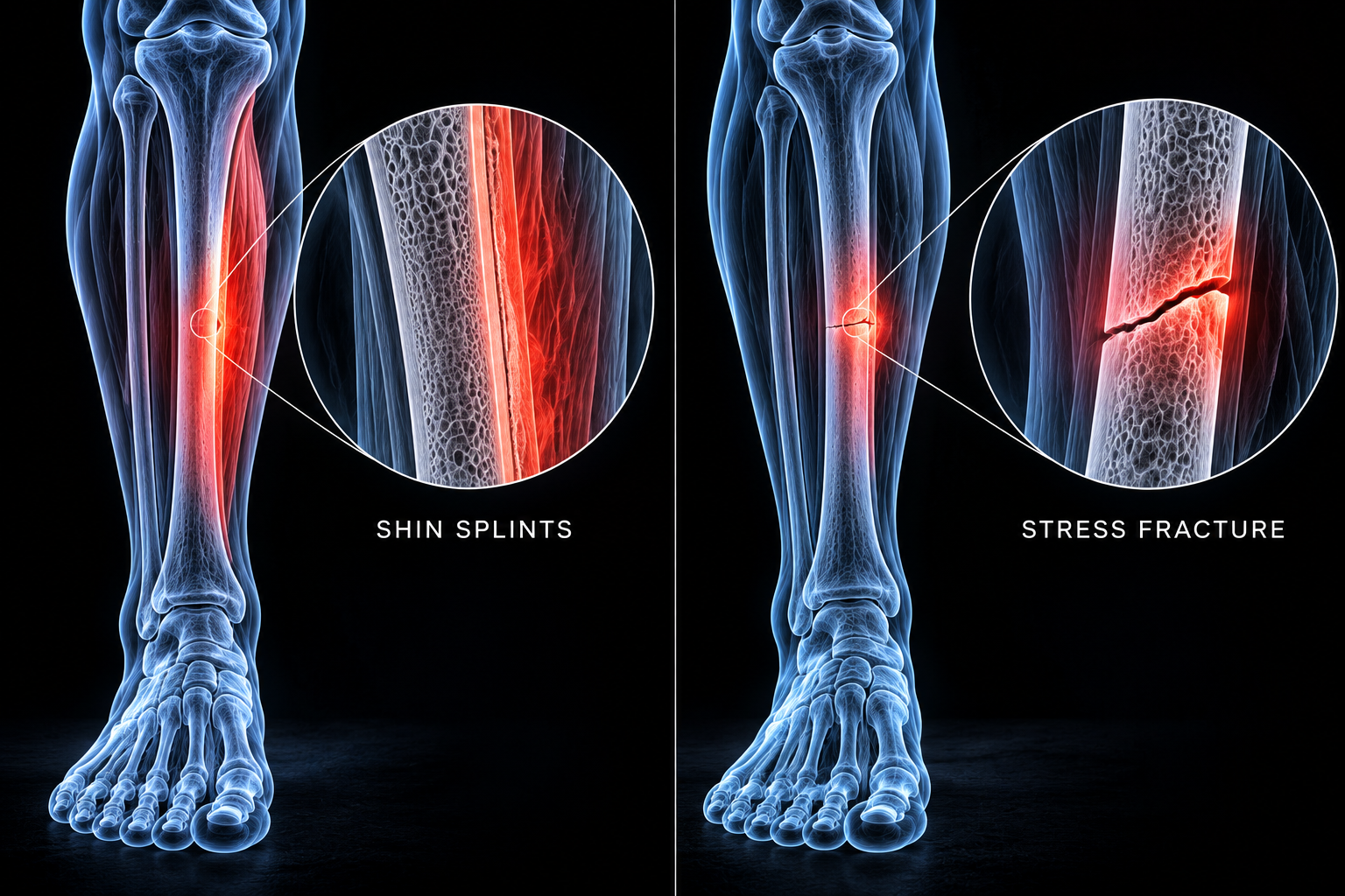
Shin pain during training is one of the most common complaints in running, soccer, basketball, and any sport that involves repeated ground contact. Most athletes call it "shin splints" and keep going — either because the pain is manageable or because they don't know what else to do. The problem is that shin splints and tibial stress fractures sit on the same spectrum of bone stress injury, and the two conditions look similar early on. But they are managed very differently, and continuing to train through a stress fracture that has been misidentified as shin splints is one of the most reliable ways to turn a six-week recovery into a six-month one.
Understanding the difference isn't just academic. The location of the pain, when it appears in a training session, whether it resolves quickly with rest, and where exactly the bone is tender are all clinically meaningful signals. At KRU Physical Therapy + Performance Lab, distinguishing between these two presentations is one of the first steps in managing an athlete with lower leg pain — because the treatment decisions that follow depend entirely on getting that distinction right.
01 | What They Are and Why They're Confused
Shin splints — the clinical term is medial tibial stress syndrome, or MTSS — is an overuse injury characterized by diffuse pain along the inner border of the lower two-thirds of the tibia. The pain comes from cumulative mechanical stress on the bone and the connective tissue that attaches to it, producing a periosteal stress reaction without a discrete fracture line.
A tibial stress fracture is a structural failure of the bone — a crack that develops when repetitive loading exceeds the bone's capacity to remodel and repair between sessions. Stress fractures occur on a continuum: early bone stress reactions look nearly identical to MTSS on exam and can only be distinguished by imaging. A true cortical stress fracture is further along that same spectrum and carries significantly higher consequences if loading continues.
The confusion arises because both conditions:
- Develop gradually in response to a spike in training load
- Produce pain along the tibia during activity
- Often improve with short periods of rest before returning with the next training block
- May be initially negative on plain X-ray
The difference is in the specifics — and those specifics change the management completely.
02 | The Symptom Pattern: What Each Feels Like
Medial Tibial Stress Syndrome (Shin Splints)
MTSS typically presents as a dull, diffuse ache along the inner shin that comes on during activity and, in early stages, resolves quickly with rest. Athletes often notice it in the first mile of a run and then find it lessens as they warm up — only to return after stopping. As the condition progresses, the pain onset moves earlier in the session, the warmup period stops providing relief, and eventually pain persists throughout activity and into the hours afterward.
The tenderness covers a broad area — typically spread over 5 centimeters or more along the middle to lower inner border of the tibia. Pressing on that area is uncomfortable but not dramatically so. There is usually no significant swelling, no pain at rest, and no pain with normal walking in the early stages.
Tibial Stress Fracture
Stress fractures tend to produce a sharper, more localized pain that progresses faster and is less reliably relieved by warmup. Several features distinguish the symptom pattern from MTSS:
- Pain that stops activity, rather than improving once warmed up. An athlete who has to cut a run short due to increasing pain — not just discomfort — should treat this as a stress fracture until proven otherwise.
- Pain with walking or normal daily activity in more advanced cases. MTSS rarely limits walking; a significant stress fracture often does.
- Night pain or pain at rest is uncommon with MTSS and should raise suspicion for a more serious bone stress injury.
- Rapid onset following a significant training spike — a sudden increase in mileage, a new surface, or a rapid return to training after time off.
03 | The Clinical Exam: How a PT Distinguishes the Two
The single most important clinical differentiator between MTSS and a tibial stress fracture is the nature of the tenderness on palpation.
Diffuse vs. Focal Tenderness
MTSS produces nonfocal tenderness — pressure is uncomfortable across a wide span of the posteromedial tibia, typically covering the middle to lower third. A tibial stress fracture produces focal, pinpoint tenderness at a specific spot. In clinical practice, tenderness that is confined to less than 5 centimeters on the anterior or posteromedial tibia warrants imaging to rule out a fracture, particularly when combined with a history consistent with high or rapidly increased training load.
Location Matters: Posteromedial vs. Anterior
Where on the tibia the pain is located also matters clinically:
- Posteromedial tibia (inner shin): The typical location of MTSS. Stress fractures at this location — on the compression side of the bone — are considered lower-risk and tend to respond well to conservative management with activity modification and a graded return to running over 6–8 weeks.
- Anterior tibia (front of the shin): This is a high-risk stress fracture location. The anterior cortex is the tension side of the tibia — it is under tensile load with each step, which dramatically impairs healing and increases fracture risk. Anterior tibial stress fractures fail conservative management in a significant proportion of cases. Up to 70% ultimately require surgical intervention, typically intramedullary nailing. A dreaded black line visible on X-ray at this location is a sign of a chronic, poorly healing fracture that needs urgent specialist attention.
Clinical Tests
Two tests are commonly used to help differentiate the conditions at the clinical level, though neither is definitive on its own:
- Hop test: Single-leg hopping on the affected leg. Reproduction of sharp, localized pain suggests a stress fracture. The test has limited specificity — it is positive in 70–100% of confirmed stress fractures but also in roughly 45% of MTSS cases — so a positive hop test increases suspicion but does not confirm a fracture.
- Tuning fork test: A vibrating tuning fork placed over the suspected fracture site may provoke pain locally. One study found 75% sensitivity and 67% specificity for tibial stress fractures — useful as a screening tool but not diagnostic on its own.
04 | Imaging: When You Need It and What It Shows
| Imaging Type | What It Shows | Sensitivity for Stress Fracture | When to Use |
|---|---|---|---|
| X-ray | Normal in early stress injury; may show periosteal reaction, sclerosis, or fracture line in advanced cases | ~10% initially; rises to 30–70% after 3 weeks | First-line, low-cost — appropriate initial study. Negative early X-ray does not rule out fracture. |
| MRI | Bone marrow edema, periosteal edema, cortical fracture line. Grades stress injury severity (Fredericson classification). Shows surrounding soft tissue. | High sensitivity and specificity; superior for grading severity | Gold standard for bone stress injury when X-ray is negative and clinical suspicion remains. Best for staging and return-to-sport planning. |
| Bone scan (scintigraphy) | Focal uptake suggests fracture; diffuse uptake more consistent with MTSS | 74–100% sensitivity | Useful when MRI is unavailable; lower specificity and more radiation than MRI. Cannot distinguish stress reaction from infection or tumor without clinical context. |
| CT | Fracture line detail; useful for surgical planning | Lower than MRI; more radiation | Not first-line for diagnosis; used when surgical planning is needed or MRI is contraindicated. |
One important clinical note: MRI can detect bone remodeling and stress reactions in completely asymptomatic runners — one study found stress reactions on MRI in 43% of asymptomatic collegiate runners, none of whom progressed to fracture. This means MRI findings always need to be interpreted in the context of the clinical picture. The imaging tells you what's happening structurally; it doesn't make the management decision by itself.
05 | Management: How Treatment Differs
Shin Splints (MTSS)
MTSS is managed conservatively through load modification and addressing the underlying contributors. The evidence is clear that complete rest is not necessary or ideal — the goal is to reduce the total mechanical stress through the tibia to a level the bone can tolerate, then progressively rebuild capacity.
- Reduce load by temporarily substituting lower-impact activities: swimming, cycling, and pool running maintain cardiovascular fitness without the repetitive tibial impact of running.
- Address training errors: MTSS is almost always preceded by a rapid increase in mileage, surface change, or return to training after time off. The training load that caused the problem needs to be corrected before a return is attempted.
- Address biomechanical contributors: Overpronation, increased ankle plantarflexion stiffness, hip weakness, and reduced running economy are all associated with MTSS. These aren't fixed by rest — they require specific rehabilitation.
- Graded return: A structured walk-to-run progression starting when the athlete is pain-free with daily activity. The typical return timeline is 6–8 weeks for MTSS managed appropriately from early presentation.
Tibial Stress Fracture
Management depends heavily on fracture location and MRI grade.
- Low-risk (posteromedial cortex): Activity restriction, possible partial weight-bearing in severe cases, cross-training for fitness maintenance. Healing typically takes 4–12 weeks. A progressive return to running follows only after the athlete is pain-free with walking and low-impact loading.
- High-risk (anterior cortex): This requires specialist input — a sports medicine physician or orthopedic surgeon. Running must stop. A pneumatic brace may be used. Surgical consultation is appropriate earlier in the management timeline given the high rate of conservative treatment failure. Up to 70% of anterior tibial stress fractures fail conservative management and require surgery; those that are caught late carry risk of complete fracture.
06 | Risk Factors and Prevention
Both conditions share several risk factors, and understanding them is the foundation of prevention:
- Prior MTSS or stress fracture is the strongest predictor of recurrence. An athlete who has had shin splints before is significantly more likely to develop it again unless the underlying load tolerance and biomechanical issues have been directly addressed — not just rested through.
- Rapid training progression — increasing weekly mileage by more than 10% per week, adding a new training surface, or returning to full training volume after time off without a graded ramp-up is the most modifiable risk factor for both conditions.
- Bone stress injuries are more common in female athletes due to the interaction of energy availability, hormonal factors, and bone density. Athletes with irregular or absent menstrual cycles and low energy availability have significantly elevated stress fracture risk.
- Footwear and surface changes alter the mechanical loading pattern through the tibia and have been associated with MTSS onset, particularly transitions to minimalist footwear or harder training surfaces.
- Training load monitoring is the most evidence-supported prevention strategy for bone stress injuries. The internal to external load ratio — how hard the athlete reports working relative to what the session demanded — is a useful early signal for accumulating stress before symptoms emerge.
- Shin pain that worsens across a training block rather than improving is a clinical signal that warrants evaluation before continuing to train through it. Athletes tend to self-manage and underreport; a culture of early disclosure and evaluation protects against the progression from stress reaction to fracture.
- Early-season and preseason are the highest-risk windows, particularly for athletes returning from an off-season with reduced training volume. The bone adaptation that underpins stress fracture resistance develops over time — athletes who return to full training volume without an appropriate buildup are at the highest risk in the first 4–6 weeks of the new season.
Shin Pain That Won't Go Away? Get It Assessed Before It Becomes Something Worse.
At KRU Physical Therapy + Performance Lab, we assess bone stress injuries across the full clinical spectrum — from early MTSS through stress fracture screening — and build return-to-sport progressions based on what the tissue can actually handle. Two locations across South Florida, plus telehealth worldwide.
References
- Bhatt D, et al. Shin Pain in Athletes. Aust J Gen Pract. 2023;52(11). (MTSS vs. anterior tibial stress fracture differentiation; epidemiology, exam findings, management protocols.)
- Sanderlin BW, Raspa RF. Common stress fractures. Am Fam Physician. 2003;68(8):1527–1532. Updated 2011. (Tibial stress fracture prevalence 23.6% of all stress fractures; focal vs. diffuse tenderness; imaging sensitivity; hop and tuning fork test data.)
- Medial Tibial Stress Syndrome: A Scoping Review of Epidemiology, Biomechanics, and Risk Factors. PMC. 2025. (Incidence 4–17% in athletic populations; risk factors including prior MTSS, female sex, pronation, training load.)
- Highlights of Medial Tibial Stress Syndrome in Military Recruits: A Narrative Review. PubMed. 2024. (MTSS epidemiology in high-load populations.)
- Fredericson M, et al. Tibial stress reaction in runners: Correlation of clinical symptoms and scintigraphy with a new magnetic resonance imaging grading system. Am J Sports Med. 1995. (Fredericson MRI classification; asymptomatic stress reactions in 43% of collegiate runners.)
- Boden BP, Osbahr DC. High-risk stress fractures: evaluation and treatment. J Am Acad Orthop Surg. 2000. (Anterior tibial stress fracture high-risk classification; surgical intervention rates.)